2Legal Medicine Research Center, Legal Medicine Organization, Tehran, Iran
3Mashhad Legal Medicine Research Center, Legal Medicine Organization, Mashhad, Iran
4Department of Public Health, School of Health, Shahid Beheshti University of Medical Sciences, Tehran, Iran
Knowledge about the variations in liver is essential for surgeons and radiologists, so as to prevent wrong interpretation and diagnosis. So far, no data are available on the standard characteristics of normal liver in the Iranian population. Hence, the aim of this study was to evaluate the standard data of a normal liver including weight, length, width, thickness and lobes variations of the liver of Iranians. This cross sectional study was perfomed on 600 cadavers in Mashhad Legal Medicine Center in 2016. After obtaining demographic characteristics of cadavers, the weight, length, width, thickness and variations of the liver lobes were evaluated. Data were analyzed using SPSS software. The mean values of the liver length, width, thickness, weight, and index of the liver were 23.56 ± 16.37 cm, 14.18 ± 3.49 cm, 6.53 ± 1.71cm, 1357.59 ± 381.5 g and 25.44 ± 47.72 respectively. The average length of the portal vein was 5.73±1.56 cm, while the average diameter of the portal vein was 8.90 ± 2.23 mm. Based on Netter′s anatomy, most livers (56.7%) were Type III (saddle like liver) and the lowest number of livers (2.2%) were Type II (left liver lobe atrophy). Although, the dimensions and weight of the liver was more in men than women, but except for the portal vein diameter, there was no statistically significant difference between the sexes in other values. There were significant differences between the dimensions of the liver with age, body weight and BMI. Investigation of liver morphological characteristics is useful for surgeons as well as anatomists. Besides, it includes the standard data of anthropometry in the Iranian population.
Keywords: Human liver; Cadaver; Gross anatomy; Morphometry
Liver is the largest gland in the body, mainly located in the epigastric region and right hypochondor, and is widely spread to the right hypochondor area. 1 The liver plays important functions such as detoxification, blood filtration, fat storage, as well as the production of bile and plasma proteins. 2 The liver has diaphragmatic, anterior and visceral surfaces. The liver is divided into right and left lobes by a line which passes through the gallbladder and inferior vena cava. The quadrate and caudate lobes of the liver can be seen in the visceral surface. 1 The liver bud appears at the end of the anterior gut endoderm in about the twenty-fifth day and erythropoiesis starts in the seventh month of the fetal period. During growth, the liver bud divides into two parts; the large part of the bud makes the liver, while the hepatic duct and small part of the bud makes the gallbladder and cystic duct. 3 Impaired development of the left lobe of the liver may lead to gastric volvulus, while problems in the development of the right lobe of the liver leads to portal vein hypertension. 4 Besides, the accessory lobe of the liver, especially in small size is important because it may be mistaken for a lymph nodule or cause of bleeding during surgery. 4 Information on the accessory fissure of the liver is also important because it is distinguished from the large and pathological fissures. Accessory fissure can mimic liver pathological nodes in CT images. It may also accumulate haemoperitoneum or ascite in the accessory fissures.5,6 Hence, knowdlege regarding the variations in liver lobes is essential for surgeons and radiologists, so as to prevent wrong interpretation and diagnosis.
Many factors such as age, sex, race, physical activity, nutrition, and health status affect the size of the liver and various other organs. 7 In Gray′s Anatomy, the liver length, width and thickness are 21 to 22.5 cm, 15 to 17.5 cm and 10 to 12.5 cm, respectively. 1 It has also been noted that the weight of the liver is about 1.5 kg and the portal vein is 5 to 8 cm in length and 1 to 3 cm in diameter. 1
A study in America showed that 56.4% of livers have different kinds of variations, particularly the liver with lingual process. 7 In the US, liver weight in males was 1561 g in men 8 and 1288 g in women. 9 A study on the African continent reported the average weight of the liver to be 1516 g in men and 1333 g in women. Among Nigerian population, in men, the average length of liver was 26.4 cm, width 15.7 cm and thickness 6 cm, as compared to women with average length of 25.4 cm, width 15 cm and thickness 5.5 cm. 10
A study in India showed that the liver lobe is 14.6%, left lobe atrophy 4.8% and accessory fissure is 12.1%. 11 In Indian men, liver weight was between 1210 to 1426 g and in women 1092 to 1292 g. 12-14 In Thai population, liver weight was found to be between 1252 to 1439 g in men and 1106 to 1214 g in women. 15, 16 The weight of this gland was between 1157 to 1447 g in Chinese men and 1029 to 1379 g in Chinese women.17 During liver autopsies in Iran, two studies were conducted. One study reported variations of bile duct in 8.93% of livers obtained from 50 cadavers in Kerman city 18 while another noted liver weight, length and diameter of portal vein in Tehran was 1453 g, 8.3 mm and 11.6 mm respectively. 18, 19
In Western countries, texts are based on anthropometric characteristics. According to this survey, there are few studies with low sample size about the standard data of normal liver in the Iranian population. Hence, the aim of this study was to investigate the standard data of a normal liver such as length, width, weight, thickness, variation of lobes and portal vein in the Iranian population.
This crosssectional study was undertaken on 600 cadavers (124 female, 476 male), referred to the dissection hall of the Forensic Medicine Organization, Razavi Khorasan province, from February 2016 to June 2016. This research was approved by the Ethics Research Committee of Mashhad Legal Medicine Organization.
Type of death was fall from a height, accident, and stab wound. Time between death and determining parameters was not more than 24 hours. Fresh Iranian cadavers with no history of poisoning, alcohol, smoking, or drug abuse; no sign of decomposition; and no evidence of trauma or abnormality of the liver were included in the study. Cadavers with cirrhotic liver, fatty liver, or any disease that increase or reduce the liver size were excluded from the study.
Demographic data, including gender, age, and body weight and height were recorded. The index was calculated as liver weight divided by body weight. Body mass index (BMI) was also calculated as weight (kg)/ height (m2).
Six hundred cadavers were divided into 10 different age groups: Group I (0- 9 years), Group II (10- 19 years), Group III (20- 29 years), Group IV (30- 39 years), Group V (40- 49 years), Group VI (50- 59 years), Group VII (60- 69 years), Group VII (70- 79 years), Group IX (80-89 years), and Group X (90- 99 years).
The thorax was opened by a midline incision, and the liver was washed with tap water. The length of the liver was measured from the base to the apex using a vernier caliper. Caliper calibration performed previously based on ISO guidelines. The greatest distance between the anterior and posterior surfaces of the liver was considered to be its thickness. The liver’s weight was also measured with the help of an electronic weighing scale.
The portal size was measured using a standard vernier caliper. Measurements for all cadavers were performed by an expert anatomist. Photographs were taken using a Canon digital camera. The classification of liver types was performed as follows: 7
Type I: small left lobe with deep costal impression;
Type II: atrophy left lobe;
Type III: saddle like liver with large left lobe;
Type IV: liver with tongue like process;
Type V: deep renal impression; and
Type VI: diaphragmatic fissures.
Six hundred Iranian cadavers (124 females and 476 males) with a mean age of 42.92 ± 19.43 years were included in the study. The values obtained for height ranged between 16 and 196 cm. The weight of the cadavers ranged from 2 to 96 kg. The mean body mass index was 23.11±4.44 kg/m2.
The mean length of the liver was 23.56 cm (range, 3 to 33 cm). The average width of the liver measured 14.18 cm. The minimum weight of the liver was 48 grams, and its maximum Data were presented as mean ± standard deviations. p values less than 0.05 were considered significant. Kolmogorov-Smirnov test used for evaluation of the data normality. The association between anthropometric data and morphometric value of the liver were evaluated using the Pearson’s correlation. Data were analyzed using SPSS 20.0 software, independent sample t-tests and analysis of variance were perfomed.
Demographic data including age sex, and height and weight of the study population were collected (table-I).
|
Age groups |
Age (years) |
Gender (female/male) |
Height (cm) |
Weight (kg) |
|
<10 |
2.83±2.37 |
11/15 |
95.57±29.91 |
11.51±11.54 |
|
10-19 |
15.93±2.46 |
9/21 |
163.23±7.47 |
58.90±11.24 |
|
20-29 |
24.61±2.78 |
24/89 |
164.37±15.67 |
62.68±10.69 |
|
30-39 |
34.50±2.65 |
22/82 |
165.11±12.31 |
65.84±10.02 |
|
40-49 |
44.48±2.78 |
15/87 |
164.10±15.94 |
65.93±8.01 |
|
50-59 |
54.37±2.54 |
17/68 |
165.14±18.78 |
65.61±9.68 |
|
60-69 |
64.89±2.61 |
1/16 |
151.90±34.72 |
66.42±8.46 |
|
70-79 |
73.43±2.56 |
7/23 |
165.76±7.25 |
66.06±8.60 |
|
80-89 |
83.05±2.63 |
2/15 |
165.11±6.25 |
64.00±11.05 |
|
90-99 |
90.00±0.00 |
1/0 |
168.00±0.00 |
59.00±0.00 |
Values are presented as mean± SD or number.
weight was 2666 grams. The length of portal vein ranged between 0.5 and 8.5 cm, with a mean of 5.73 ± 1.56 cm. The diameter of portal vein was 8.90 ± 2.23 mm (range, 1 to 15). The index of the liver varied from 0.5 to 76, with a mean value of 25.44.
Age groups |
Length(cm) |
Width(cm) |
Thickness(cm) |
Weight(g) |
Index |
length of portal vein(cm) |
Diameter of portal |
<10 |
21.26±78.36 |
3.82±2.41 |
1.86±1.28 |
291.61±217.56 |
34.96±29.58 |
1.94±1.27 |
3.05±1.95 |
10-19 |
22.70±4.64* |
13.43±2.86* |
6.78±1.12* |
1360.93±451.19* |
24.34±10.17* |
6.33±1.34* |
9.05±2.41* |
20-29 |
24.07±3.35 |
15.05±2.29 |
6.73±1.60 |
1403.22±287.83 |
34.00±99.56 |
5.74±1.47 |
8.94±2.25 |
30-39 |
24.05±3.14 |
14.91±2.69 |
6.70±1.39 |
1424.35±281.78 |
25.96±44.45 |
5.71±1.19 |
9.38±1.93 |
40-49 |
23.78±3.09 |
14.78±2.73 |
6.70±1.51 |
1400.09±343.29 |
21.40±5.50 |
6.04±1.40 |
9.13±1.61 |
50-59 |
23.60±3.76 |
14.63±2.99 |
6.75±1.48 |
1443.91±317.82 |
22.44±5.63 |
5.90±1.18 |
8.99±1.53 |
60-69 |
23.27±3.47 |
14.39±2.83 |
6.89±1.04 |
1408.53±279.03 |
21.61±5.53 |
5.95±1.31 |
9.53±1.56 |
70-79 |
23.20±3.80 |
14.13±2.86 |
6.81±1.32 |
1370.10±339.52 |
21.17±6.36 |
6.16±1.40 |
8.81±1.87 |
80-89 |
22.94±3.61 |
14.02±2.51 |
6.35±1.27 |
1275.47±157.68 |
20.35±3.71 |
5.64±1.53 |
9.41±1.46 |
90-99 |
24.00±0.00 |
16.00±0.00 |
8.00±0.00 |
1534.00±0.00 |
26.00±0.00 |
6.00±0.00 |
9.00±0.00 |
Values are expressed as mean± SD. Comparison between groups was made using ANOVA and Tukey test. *p<0.05 compared to group A within column
The smallest width of the liver was found in the first of life, while the greatest width of the liver was seen in the ninth decade. The weight of the liver was heaviest in cadavers 90–99 years old, while it was lightest in cadavers 0 - 9 years old. The index of the liver was the largest in Group I, and least in Group IX.
The morphometric characteristics of the liver of cadavers in males and females were assessed. The diameter of the portal vein was significantly higher in males than females (table III) but it was not statistically significant (p>0.05).
Gender |
|||
Morphometric characterizes |
Female |
Male |
p value |
Length (cm) |
22.19±5.73 |
23.92±18.15 |
0.29 |
Width (cm) |
13.65±3.85 |
14.31±3.38 |
0.06 |
thickness(cm) |
6.38±2.00 |
6.57±1.63 |
0.32 |
Weight (g) |
1312.21±463.38 |
1368.35±356.44 |
0.20 |
index |
24.04±14.55 |
25.81±53.12 |
0.71 |
length of portal vein (cm) |
5.49±1.68 |
5.79±1.52 |
0.05 |
diameter of portal vein (mm) |
8.47±2.49 |
9.0179±2.15 |
0.02 |
Values are presented as mean± SD. Independent samples t-test was used to compare values
The correlation of morphometric data of the liver with the anthropometric values are shown in table IV.
Morphological parameters |
Age |
Height |
Body weight |
BMI |
|
Length (cm) |
Pearson Correlation |
0.002 |
0.000 |
0.01 |
-0.42 |
Width (cm) |
Pearson Correlation |
0.24** |
0.0 |
0.48** |
0.36** |
Thickness (cm) |
Pearson Correlation |
0.27** |
0.04 |
0.45** |
-0.12 |
Index |
Pearson Correlation |
-0.72 |
-0.30 |
-0.33** |
-0.28** |
Weight (g) |
Pearson Correlation |
0.25** |
0.01 |
0.48** |
0.11** |
length of portal vein(mm) |
Pearson Correlation |
0.13** |
0.04 |
0.41** |
0.31** |
Diameter of portal vein(mm) |
Pearson Correlation |
0.27** |
0.09* |
0.45** |
0.34** |
Correlations were assessed using Pearson correlation coefficients.** Significant in p value 0.01. * Significant in p value 0.05
The width and weigth of liver were strongly correlated with body weight, BMI, and age. A significant correlations were evident between the value of demographic data and portal vein size (p<0.05). The correlation was significant for index of liver with body weight and BMI. In adition, the thickness of liver was strongly correlated with age and body weight.
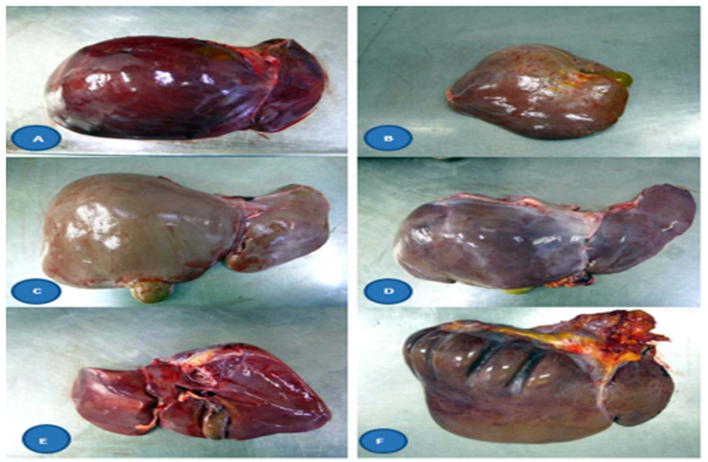
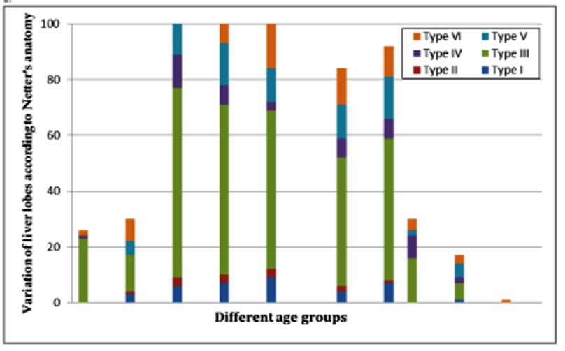
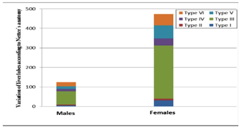
Congenital or acquired variations in these samples were to be found. The results of classification of liver types according to Netter′s anatomy are shown in Figure 1- 3. In overall, type I (small left lobe with deep costal impression) was observed in 37 livers (6.2%). Type II (atrophy left lobe) were seen in 14 livers (2.6%). Saddle like liver with large left lobe and liver with tongue like process were in 341 (56.7%) and 47 livers (7.8%), respectively. Liver type V and VI were found in 13.5% and 13.2% of cadavers respectively.

Figure 1: A. Netter′s Type I liver showing small left lobe. B. Type II liver showing atrophy left lobe. C. Type III liver showing: Saddle like liver with large left lobe. D. Type IV liver showing liver with tongue like process. E.Type V liver showing deep renal impression, F. Type VI liver showing diaphragmatic fissures

Figure 2: The incidence of variation of liver lobes of Iranian cadavers in different age groups

Figure 3: The incidence of variation of liver lobes of Iranian cadavers in males and females.
The mean values of the liver length were 23.56 ± 16.37 cm, width 14.18 ± 3.49 cm, and thickness 6.53 ± 1.71 cm. Although, the liver size was more in men than women, it was not statistically significant. Many factors such as age, sex, race, physical activity, nutrition, and health status affect the size of the liver. 7 Besides, development of liver may affect variation of liver lobes especially presence of accessory lobes. In Gray′s Anatomy, liver length was reported to be between 21 to 22.5 cm, width 15 to 17.5 cm and a thickness of 10 to 12.5 cm. 1 The mean length of the liver in Nigerian men was 26.4 cm, width 15.7 cm and thickness 6 cm; but it was not significant as compared to Nigerian women, with an average length of 25.4 cm, width 15 cm and thickness 5.5 cm. 10
In this study, the average weight of the liver was 1368 g for men and 1312 g for women. There was a significant relationship between liver weight and age as well as liver weight and BMI. In both sexes, there was no significant difference in liver weight. Due to more haemopoitic activity in men than women, higher weight was observed in men as compared to women. According to Grant anatomy, the liver weight in adult females varies from 1200 to 1400 g and 1400 to 1800 g in adult males. 21 In Gray′s Anatomy and Moore, liver weight of 1500 g in adults was noted (2.5% of body weight). 1, 22 Liver weights in American men were reported to be 1561 g and 1288 g in American women. 8, 9 Liver weights are listed as 1516 g in Nigerian men and 1333 g in Nigerian women. 10 In India, liver weight was 1210 to 1426 g in men and 1092 to 1292 in women. 12, 13, 14 For Korean men, liver weight was reported as 1650.40 and 1344.10 g for Korean women. There was no association between liver weights with age in both sexes, but there was an association between height, body weight and liver weight. 23 Liver weight was 1157 to 1447 g in Chinese men and 1029 to 1379 g for Chinese women. 17 The average weight of the liver in 40 autopsies in Tehran was 1453 g. 19
Based on Netter′s anatomy, most livers (56.7%) were Type III (saddle like liver) and the lowest livers (2.2%) were Type II (left liver lobe atrophy). Nagato in Brazil reported that 42.6% of livers were normal while 56.4% had liver variations such that most of them were liver with a tongue-like process. 7 The results of Patil′s study on 50 cadavers showed that liver type I, IV, V and VI, were observed in 2% of cases. 4 Type III liver was found in 10% of cases while there was none in type II liver.
The average length of the portal vein was 5.73±1.56 cm and the average diameter of the portal vein was 8.90 ± 2.23 mm. In anatomy text books, the length and diameter of the portal venous has been reported to be between 5 to 8 cm and 10 to 30 mm, respectively. 1 Bannazadeh et al reported that the length and diameter of portal vein in 40 autopsies in Iran to be 8.3 cm and 11.6 mm respectively. 19
In this study, the number of males was more than females; this may lead to sampling bias. Based on the search, there has been no research on the standard data of the liver in the Iranian population, and this is the first report and strength of this study. The weakness of this study was the unavailability of data about the variations of the bile ducts and liver artery. However, for the best results, further studies including variations of the bile ducts and liver artery are recommended with larger and nationally represntative sample size.
The author would like to thank the Mashhad Legal Medicine Organization for their help and financial support.
References
- Dark RL, Vogle AW, Mithell AWM. Gray’s anatomy, in Alimentary System, Churchill Livingstone, 3th ed. New York, NY, USA; 2015. P 328-32.
- Mescher AL. Junqueira’s Basic Histology. 13th ed. Singapore: MC Graw Hill Education; 2013. P 329-40.
- Sadler TW. Langman′s medical embryology. 13th ed. Philadelphia: Wolters Kluwer; 2015. P 236-7.
- Patil S, Sethi M, Kakar S. Morphological study of human liver and its surgical importance. Int J Anat Res 2014; 2:310-14.
- Mehta V, Arora J, Manik P, Suri RK, Rath G. Clinico-anatomical aspects of accessory fissures obscuring the normal hepatic morphology. Clin Ter. 2010; 161: 259-60.
- Auh YH, Rubenstein WA, Zirinsky K, Kneeland JB, Pardes JC, Engel IA et al. Accessory fissures of the liver: CT and sonographic appearance. Am J Roentgenol. 1984; 143:565-72.
- Nagato AC, Silva MAS, Trajano ETL, Alves JN, A Bandeira ACB, Ferreira TA et al. Quantitative and morphological analyses of different types of human liver. J Morphol. Sci 2011; 28: 275-9
- Molina DK, DiMaio VJ. Normal organ weights in men: part II-the brain, lungs, liver, spleen, and kidneys. Am J Forensic Med Pathol. 2012; 33:368-72.
- Molina DK and DiMaio VJ. Normal Organ Weights in Women: Part II-The Brain, Lungs, Liver, Spleen, and Kidneys. Am J Forensic Med Pathol. 2015; 36:182-7.
- Emue EB, Amaza DS, Sambo N, Hamman LL, Nggada HA, Tarfa M. A Study of the Normal Morphological Variations of Adult Nigerian Human Cadaveric Liver. JDMS; 2013; 3:21-3.
- Muktyaz H, Nema U, Rakesh G, Arun KS. Morphological variations of liver lobes and its clinical significance in north Indian population. GJMMS. 2013; 1:1-5.
- Prakash C, Deopa D, Thakkar HK. Study of Internal Organ Weight and Its Correlation to Body Weight in Kumaon Region of Uttarakhand . J Indian Acad Forensic Med. 2013; 35: 29- 32.
- Singh D, Bansal YS, Sreenivas M, Pandey AN, Tyagi S. Weights of human organs at autopsy in chandigarh zone of north-west India. JIAFM. 2004; 26: 97-99.
- Tanna JA, Patel PN, Kalele SD. Relation between Organ Weights and Body Weight in Adult Population of Bhavnagar Region- A Post-Mortem Study. J Indian Acad Forensic Med. 2011; 33: 57-9.
- Narongchai P and Narongchai S. Study of the normal internal organ weights in Thai population. J Med Assoc Thai. 2008; 91:747-53.
- Chirachariyavej T,Ouyswat K, Sanggarnjanavanich S, Tiensuwan M, Peonim V, Sirikulchayanonta V. Normal Internal Organ Weight of Thai Adults Correlated to Body Length and Body Weight. J Med Assoc Thai. 2006; 89: 1702-12.
- In Human Liver Location and morphology. [Online] cited 2010. Available from: http://www.ganbling.com.cn/YLX.H TM
- Eftekhar SH, Shams Ara A, Jamalizadeh M. A study of the anatomic variations in extrahepatic bile ducts in 50 adults refereed to Kerman Forensic Medicine Organization. Iranian Society of Anatomical Sciences. 2013; 10: 57-62.
- Bannazadeh H, Salehian MT, Baharestani B. Liver Weight and the Dimensions of its Vessels and Biliary Ducts: A Study on 40 Iranian Cadavers. The Archives of Iranian Medicine Journal. 2000; 3: 1-3.
- Mohammadi S, Hedjazi A, Sajjadian M, Kebriaei SMM, Naser Ghrobi N, Yaghmaei A, Moghadam MD, Mohammadi M. Morphometric Study of Spleen in Iranian Cadavers from Razavi-Khorasan Province. Bangladesh Med Res Counc Bull 2016; 42: 61-66
- Williams L. Grant’s Method of Anatomy. 15th ed. Baltimore: Williams and Wilkins; 1995. P 1796.
- Moore KL, Dally AF. Clinically oriented anatomy. 5th ed. Philadelphia: Lippincott Williams & Wilkins; 2006. P 289.
- Y, Kim D, Cho SY, Kim MH,Yang KM, Lee HY et al. Statistical Analysis for Organ Weights in Korean Adult Autopsies. The Korean J Anat. 2009; 42: 219-24.